Health & Fitness
New 3-D Imaging Tool Used To Detect Prostate Cancer
Urologists at Northside Hospital are using a combination of MRI and ultrasound to catch the cancer earlier and track its progress.


SANDY SPRINGS, GA -- Urologists at Northside Hospital have a new tool they can use to help detect and biopsy prostate cancer.
These doctors are now using a combination of magnetic resonance imaging and ultrasound with computer-aided detection to produce real-time 3-D images of the prostate to see cancer earlier and better track its progression, the hospital said Wednesday.
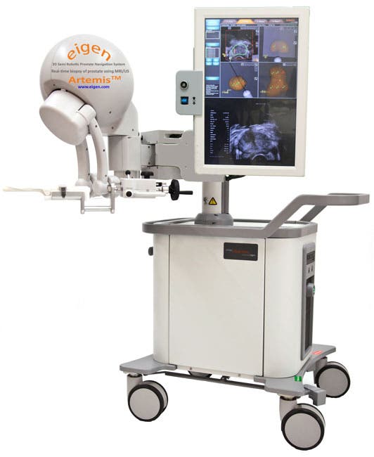
Artemis with ProFuse Bx is a 3-D ultrasound-guided prostate biopsy platform from Eigen.
Find out what's happening in Sandy Springsfor free with the latest updates from Patch.
The technology allows doctors to see 3-D images of the prostate and guide the needle during biopsy, while mapping the precise location of the tumor so that they can return to it later for follow up.
Northside Hospital is the first in metro Atlanta to acquire the Artemis system, it stated.
Find out what's happening in Sandy Springsfor free with the latest updates from Patch.
Approximately 5,570 men in Georgia will be diagnosed with prostate cancer this year, according to the American Cancer Society. Next to skin cancer, prostate cancer is the most frequently diagnosed cancer in men, the organization states.
Early detection is key in successfully treating many cancers. However, prostate cancer can be slow growing and take years to develop. Men diagnosed with the disease usually have time to consider all available treatment options, gather additional opinions and, with the help of their doctor, decide on which option is best for them.
“Still, new methods are needed to help physicians improve disease staging, select the most appropriate treatment and provide the best long-term follow-up for patients,” said Dr. Mark Haber, urologist, Georgia Urology.
The most common tools historically used to evaluate prostate cancer tumors and growth include prostate ultrasound, MRI and biopsy. The problem with each of these is, when used separately, they have limitations. Ultrasound only provides two-dimensional images and the MRI is a high-powered magnet, which prevents the use of needles during the procedure.
Biopsy alone does not give doctors a full picture of the cancer, leaving doctors basically blind and at risk of having to stick the patient multiple times.
However, when these modalities are combined, the result is a more accurate diagnosis along with better information regarding the exact location and extent of the disease, providing information that helps doctors and patients both make a smart, individualized choice about prostate cancer management or treatment.
“MRI technology and specifically, fusion biopsy, provides a more personalized and tailored approach to local prostate cancer treatment options,” said Dr. Vahan Kassabian, urologist and medical director of Georgia Urology. “This allows us to minimize potential urinary and sexual side effects.”
Every case of prostate cancer is different and based on the patient’s age, stage of the disease and their doctor’s advice, treatment options will vary.
Beginning at age 50, men at average risk (no family history) for developing prostate cancer should begin to discuss the pros and cons of screening with their doctor. Men at high risk for developing prostate cancer should begin discussing screening even sooner.
For more information about prostate cancer care at Northside Hospital, visit its website.
Image via Northside Hospital
Get more local news delivered straight to your inbox. Sign up for free Patch newsletters and alerts.