Schools
Montgomery County Community College: New Anatomage Tables Unveiled For Virtual Anatomy Exploration
Students studying health science at Montgomery County Community College this fall will get to use new, groundbreaking technology to stud ...
Eric Devlin
September 13, 2021
Find out what's happening in Ardmore-Merion-Wynnewoodfor free with the latest updates from Patch.
Students studying health science at Montgomery County Community College this fall
will get to use new, groundbreaking technology to study human anatomy and physiology.
 MCCC purchased two Anatomage Tables this past spring for students and faculty to incorporate in their study of
the body. The Anatomage Table is the most technologically advanced 3D anatomy visualization
and virtual dissection tool for anatomy and physiology education, according to its
website.
Find out what's happening in Ardmore-Merion-Wynnewoodfor free with the latest updates from Patch.
Students using the machine will soon be able to peel back and explore each layer of
a digitally recreated body, starting with the skin and musculature, down to the circulatory
and central nervous systems and everything in between.
“The Anatomage Table was developed to provide real-life dissection of a human cadaver
without having a human cadaver,” said Cheryl DiLanzo, Dean of Health Sciences. “By
using this table, students will be able to understand spatial anatomy which is crucial
in health care and STEM.”
Health science courses like anatomy, physiology, biology, nursing, dental hygiene
and other programs will likely heavily incorporate the use of the machine into their
work, she said.
“There isn’t any program that wouldn’t be able to use it for something” said DiLanzo.
“It looks like a regular table, but it’s really incredible.”
 The two tables are available for use separately at Blue Bell and Pottstown Campuses.
They were purchased using a Carl D. Perkins grant awarded by State of Pennsylvania
Department of Education, for funding technology that furthers the goals of Career
Technical Education students for jobs of the future.
“It is a very cool thing to able to get on both campuses,” said DiLanzo, adding faculty
have already begun to rave about the machine. “Faculty can demonstrate anatomy that
they are teaching in class and highlight specific areas such as vasculature, bones,
and organs very easily. The table may be used in person, or students can also learn
remotely while the faculty are teaching at the table. Additionally, faculty can add
pathologies to the cadaver, so that students gain a better understanding of what medical
conditions may look like in a human body. Faculty can incorporate case studies, labeling,
quizzes and supplemental instruction for student.”
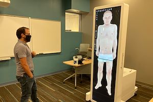
The approximately 7-feet-by-2-feet-by-3-feet machine incorporates two touchscreen
computer monitors connected to a Windows 10 operating system to project a three-dimensionally
rendered image to manipulate. The images are based on the cadavers of four deceased
individuals whose bodies were donated to science and digitally scanned. The program
also includes CT scans of each patient, along with a case library section of the software,
where over 1000 CT/MRI scans of various different individuals with case information
are available for review, said Matthew Kilbride, Instructional Technology Simulation
Specialist.
“All of them come from The National Library of Medicine’s Visible Human Project,” said Kilbride, which features complete, anatomically detailed, three-dimensional
representations of a human male body and a human female body. “There’s a male and
female Asian cadaver and there’s a male and female Caucasian cadaver. The two Caucasians
are from the United States’ Visible Human Project, and the two Asians from Korea’s,
which is labeled as Visible Korean Human.”
The approximately 300-pound machine can be converted vertically and horizontally and
incorporated for lectures, presentations and/or small, group work situations. Within
the software users can manipulate the 3D image to zoom in and out and rotate the body
as needed. Different customizable menus allow users to focus on certain sections of
the body, such as the head and neck or even just the frontal section of the skull,
for example.
“Each structure and category, you’re able to put in a label in the three-dimensional
space,” said Kilbride. “You can also add annotations. So you can create your own custom
views.”
DiLanzo credited Kilbride for helping bring everyone up to speed on how to use the
machine.
“I can’t sing his praises enough,” she said. “He knows that table. The table has so
many uses. He conducted virtual and in person trainings. He came on board and really
took this as his project and spearheaded that for us.”
For more information about the Anatomage Table, visit its website.
This press release was produced by the Montgomery County Community College. The views expressed here are the author’s own.