Neighbor News
What Are Cytokine Measurement Assays?
Expand your knowledge of cytokine measurement assays with explanations, tips, and tricks from a Seattle-based research laboratory.
Scientists use cytokine measurement assays to identify cell-signaling proteins that tell the body's immune system to initiate an immune response against an invading pathogen. These assays can help researchers understand how our immune systems react to viruses and bacteria that cause us harm.
When it comes to immunology, researchers often use assays to measure these common cytokines:
- IFNγ
- TNFα
- Interleukin 1β (IL-1β)
- Interleukin 2 (IL-2)
- Interleukin 4 (IL-4)
- Interleukin 5 (IL-5)
- Interleukin 6 (IL-6)
- Interleukin 8 (IL-8)
- Interleukin 10 (IL-10)
- Interleukin 12p70 (IL-12p70)
- Interleukin 13 (IL-13)
Cytokine Measurement Assay Type 1: Collection from Culture Medium
Find out what's happening in Seattlefor free with the latest updates from Patch.
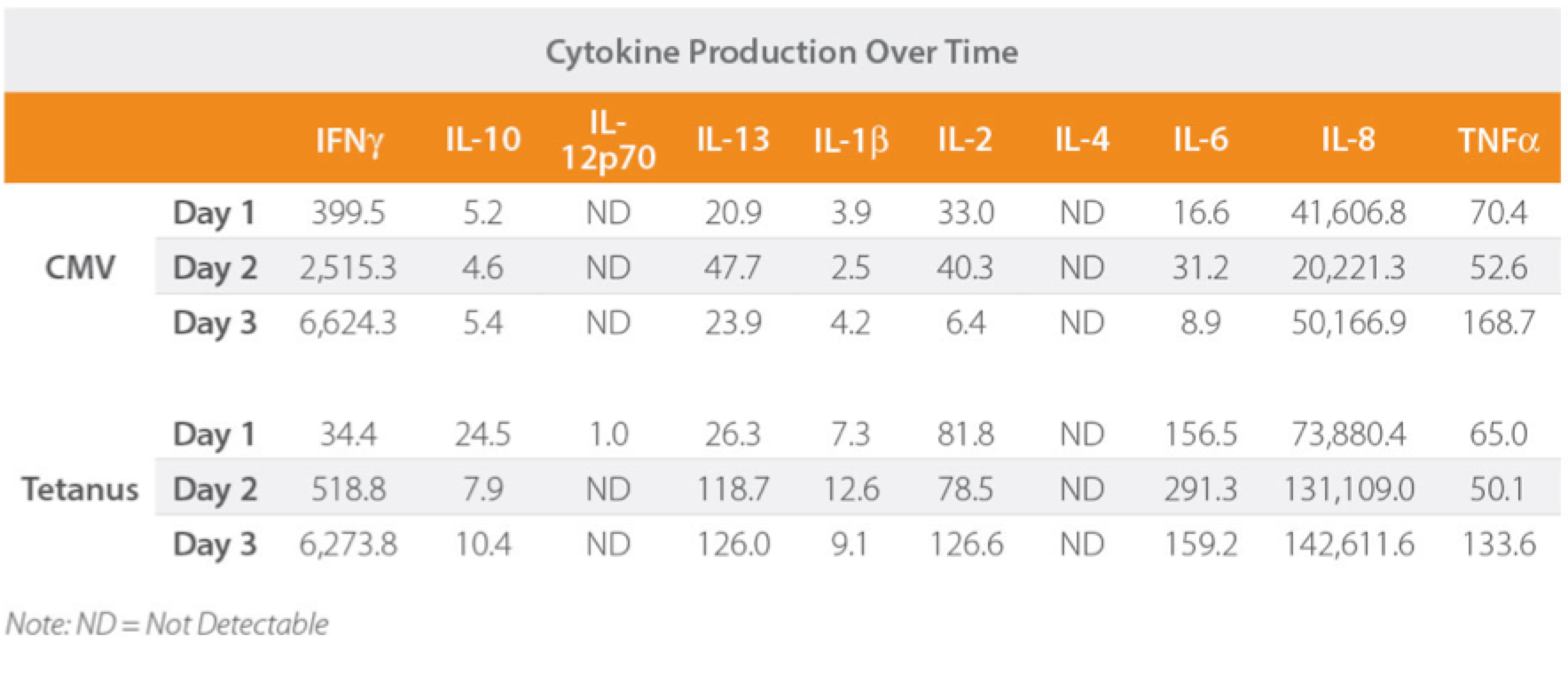
Cytokines can be measured with a single assay (ELISA) or an advanced multiplex assay (Meso Scale, Luminex) both of which have been show to be equally accurate. Both methods require an intimate understanding of the optimal measurement time for each cytokine. Culture too early and you can miss the cytokine release altogether; too late and you can get a suboptimal readout.
Keep in mind the cytokine fundamentals when analyzing your charts. For example, IL-2 may not be detectable in your readout, but remember that IL-2 is usually consumed as quickly as it is produced, so this shouldn't be surprising.
Find out what's happening in Seattlefor free with the latest updates from Patch.
This is one example of cytokine production measured over time in the presence of different stimuli.
Cytokine Measurement Assay Type 2: Intracellular Staining
Intracellular staining is a way to quantitate the reactive cells in an assay to understand which cells are involved in the immune response.
We start our intracellular cytokine staining process by incubating antigen-presenting cells overnight with peptide. The next day, we add T cells and culture again overnight. We then add brefeldin A — an antiviral used for research purposes — for the final 4 hours of the culture to prevent cytokine secretion.
We finish the assay by staining any dead cells and CD8 cells, fixing and staining for IFNγ, then permeabilizing the cells with 0.5% saponin. Finishing with the permeabilization ensures the antibody can access the inside of the cells.
You can then analyze the fluorescence to understand which cells are producing cytokines.
This post originally appeared on the Astarte Biologics blog and has been modified for Patch.