Schools
Microscopy Lab Helps Students Visualize Proteins
WCSU Department of Biology's Microscopy Lab helps students visualize proteins central to brain function and more

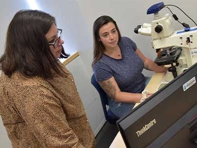
DANBURYm— Western Connecticut State University’s Department of Biology Microscopy Lab in the Midtown campus Science Building serves as a place for students and staff in the department to conduct learning and research. Unveiled in 2021, the lab was first used educationally at that time by Associate Professor of Biology Dr. Kristin Giamanco, who can’t say enough about its attributes.
Giamanco said that the hands-on experience gained will better prepare students for the professional world. Since the microscopes in the lab are widely used in the scientific community, learning how to use them properly and efficiently is “a very marketable skill.”
The microscopes and technology in the lab are sought after by researchers at WCSU who are interested in staining for proteins, genes, microstructures in cells, stem cell research and genomics. According to Biology Department Chair & Professor of Biology Dr. Theodora Pinou, there is also a tremendous interest among incoming master’s degree students in looking at marine ecology, specifically studying the accumulation of microplastics in fish, which directly affects what people eat.
Find out what's happening in Naugatuckfor free with the latest updates from Patch.
At present, the room contains five microscopes with LED lightbulbs attached for viewing and identifying components of cells being studied. In lab classes, students perform immunofluorescence to visualize protein expression through the use of the newly upgraded LED lights and filter cubes that present as red, green or blue visible light. All the microscopes are equipped with cameras, which allow still images to be taken of the samples being studied to be used for lab reports and graphics. This technique is highly valued by employers and is used in many subfields of Biology, which prepares WCSU students for an array of careers.
Specifically, students have assayed changes to the structure of cells in a rat brain after exposure to commonly used drugs such as alcohol and caffeine. Due to the technological upgrades to these systems, students instantly took photographs of their cells and measured these changes in an autonomous and hands-on way that was not possible before.
Find out what's happening in Naugatuckfor free with the latest updates from Patch.
In another project, students examined the expression of a protein that has been linked to the development of neuropsychiatric disorders such as bipolar disorder and schizophrenia. These resources support the department’s pre-health students and enhance their confidence as they get ready to enter the job market. Additionally, the department is deeply invested in supporting and training students who have been marginalized and underserved.
Giamanco’s research program has greatly benefited from the establishment of the microscopy facility in that her students have been able to create models to better study the interplay between the environments inside and outside the cell. This work better positions researchers to understand the pathology of neurological disorders like Alzheimer’s disease and decipher how addiction is regulated.
Juliet Barbieri (WCSU Class of 2022) presented her work, “Building the Perineuronal Net: Assessing the Role of Aggrecan in Molecular Assembly,” at Western Research Day last spring and was the recipient of two prestigious awards: The Provost’s Award and the Sigma Xi Student Research Award.
“In a science course, you have to learn things tangibly … you do microscopy and want a student to learn how to use the instrument, they need to have a microscope in front of them,” explained Giamanco. “The beauty of the space,” added Pinou, “is that it allows staff and students to visualize cells and learn how to stain for proteins and microstructures, which is a very important skill that employers are looking for.”