Health & Fitness
The 360° View: Detecting Breast Cancer with 3D Mammography
Preventative and Diagnostic 3D Breast Cancer Screenings Help Women at The Steeplechase Cancer Center at RWJUH Somerset
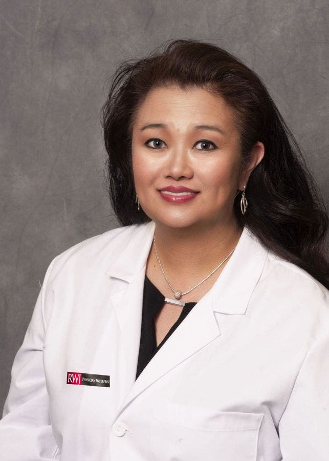
Deborah Lue, MD, a breast surgeon with RWJ Physician Enterprise Steeplechase Breast Specialists based at the Steeplechase Cancer Center at Robert Wood Johnson University Hospital Somerset
Breast cancer is the second leading cause of death in American women and is the cause of over 40,000 deaths per year. Although breast cancer affects the lives of nearly 250,000 new women each year, there are currently more than 2.8 million breast cancer survivors living within the United States. Since the late 1980s, the survival rate has continually increased due to early detection, improved treatments and heightened awareness of the disease. During National Breast Cancer Awareness Month and throughout the year, women are encouraged to take advantage of screening methods to help prevent breast cancer from taking charge of their lives.
The single most effective defense against breast cancer is screenings.
Find out what's happening in Basking Ridgefor free with the latest updates from Patch.
Women are recommended to check themselves regularly for any new lumps, masses, irritation or pain within their breasts. Breast cancer often manifests as hard, irregular masses but can be tender, soft or round. The lumps, which may or may not be large enough to feel, may be accompanied by swelling, irritation, pain or discharge from the breast and/or lymph nodes. Knowing what is “normal” for you and notifying your doctor of any hard, irregular masses can lead to early detection and early treatment.
Mammograms are important.
Find out what's happening in Basking Ridgefor free with the latest updates from Patch.
Although self-exams are an important aspect of screening for breast cancer that can be done at home, they should be paired with regular mammograms or other screening tests, because not all cancers display physical lumps or symptoms. The most common screening test, a mammography, takes two X-ray pictures of each breast. The X-ray images allow doctors to identify abnormal areas which will determine if further testing is necessary, such as a magnification views, breast ultrasound or a biopsy. We recommend that women at average risk undergo a regular screening mammography at least once a year after age 40 for the best chance of early detection. Women with a family history of breast cancer should consult with their physician to determine if earlier screenings are necessary.
3D mammography offers an opportunity for deeper analysis.
Even though mammograms are often effective in identifying cancerous masses in breasts, a 2D screening does not always provide the most comprehensive view. Many doctors are now using digital breast tomosynthesis, or 3D mammography, which uses the same X-ray technology as 2D mammography to provide several images of the breast from different angles. Doctors can view these images in single slices or as an interactive animation to examine the breast tissue for deeper analysis. By having a more comprehensive view, doctors are able to see abnormalities with greater detail that may not necessarily appear on a traditional 2D image. 3D mammograms also help decrease the need for additional follow-up imaging; lessening the financial, psychological and physical strain often associated with examining and diagnosing breast cancer.
Dense breast tissue makes finding abnormalities on mammograms harder.
For women with dense breast tissue, 3D mammography may provide an opportunity for deeper examination, but still may not detect everything. Dense breast tissue, which has a high concentration of glandular tissue compared to fatty tissue, is often difficult to examine. On X-rays, dense breast tissue and potentially cancerous abnormalities both appear white against the contrast of fatty tissue which appears black. Therefore, potentially harmful abnormalities may be hidden by healthy, dense breast tissue. At-risk women with dense breast tissue can help lower their risk for developing cancer by maintaining a healthy weight, diet and exercise regime in addition to pursuing regular screenings.
Learn more about breast care at Robert Wood Johnson University Hospital.
The Sanofi US Breast Care Program at the Steeplechase Cancer Center at Robert Wood Johnson University Hospital Somerset (RWJUH) offers the most advanced technologies to accommodate women and breast cancer patients through screening, diagnosis and treatment. A team of specially trained nurses guide patients through the program staffed by a multidisciplinary team of physicians and oncology specialists. The comprehensive program offers 3-D mammography, ultrasounds, biopsies, DEXA scanning and ImageCheckerTM computer-aided detection system to provide patients with the best care currently available.
To learn more about breast care and mammography at Robert Wood Johnson University Hospital, visit www.RWJmammo.com. To schedule an appointment for a 3D mammogram at The Sanofi US Breast Care Program at the Steeplechase Cancer Center at RWJUH Somerset, call (908) 704-3740. Interested individuals can also call (732) 253-3928 to contact Breast Care Connection at RWJUH’s New Brunswick Campus.
For more information about Robert Wood Johnson University Hospital, visit www.rwjuh.edu or call 1-888-MD-RWJUH.